anatomy and physiology of heart pdf
Anatomy and Physiology of the Heart
The heart is a vital organ that pumps blood throughout the body, delivering oxygen and nutrients to tissues and removing waste products. Understanding the anatomy and physiology of the heart is crucial for comprehending its function and the various conditions that can affect it.
Introduction
The heart, a remarkable organ, is the driving force behind our circulatory system. It acts as a tireless pump, propelling blood throughout our body, delivering oxygen and vital nutrients to every cell while simultaneously removing waste products. This intricate and essential organ, often described as the “engine” of our body, is a complex marvel of nature, its structure and function intricately intertwined.
Understanding the anatomy and physiology of the heart is paramount for comprehending its role in maintaining life and for recognizing the potential for various cardiac conditions. This knowledge provides a foundation for appreciating the delicate balance that governs heart function and the consequences of its disruption. The heart’s anatomy, encompassing its chambers, valves, and muscular structure, dictates its ability to efficiently pump blood. Meanwhile, the physiology delves into the electrical impulses that control heart rate and the intricate mechanisms of blood flow, highlighting the sophisticated choreography of the cardiac cycle.
This exploration delves into the fascinating world of the heart, unveiling its intricate design and the remarkable processes that make it possible for this vital organ to sustain life. We will journey through the chambers, valves, and intricate network of vessels that compose the heart, revealing the profound impact it has on our overall health and well-being.
Heart Chambers and Valves
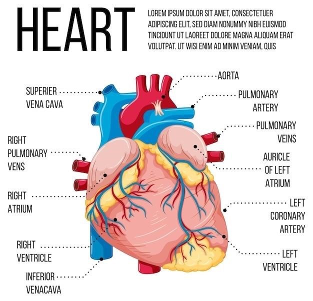
The human heart, a four-chambered organ, functions as a powerful pump, efficiently circulating blood throughout the body. These chambers, two atria and two ventricles, work in a coordinated manner to ensure continuous blood flow. The atria, acting as receiving chambers, collect blood returning from the body (right atrium) and lungs (left atrium), while the ventricles, the powerful pumping chambers, propel blood to the lungs (right ventricle) and the rest of the body (left ventricle).
Between these chambers lie heart valves, essential structures that regulate blood flow, ensuring unidirectional movement and preventing backflow. The four primary valves are the tricuspid valve (between the right atrium and right ventricle), the pulmonary valve (at the exit of the right ventricle), the mitral valve (between the left atrium and left ventricle), and the aortic valve (at the exit of the left ventricle). These valves open and close in a precise sequence, driven by pressure gradients, ensuring that blood flows in the correct direction through the heart.
The delicate interplay between the chambers and valves forms the foundation of the heart’s function. Their coordinated actions facilitate the efficient pumping of blood, delivering oxygen and nutrients to all parts of the body, a testament to the remarkable complexity of the cardiovascular system.
Cardiac Muscle
The heart, a tireless organ, relies on a specialized type of muscle tissue known as cardiac muscle to perform its crucial task of pumping blood. Unlike skeletal muscle, which is under voluntary control, cardiac muscle is involuntary, meaning it contracts rhythmically and automatically without conscious effort. This unique property ensures the heart’s continuous and efficient pumping action, essential for sustaining life.
Cardiac muscle cells, called cardiomyocytes, are highly interconnected, forming a network that allows for synchronized contraction. These cells are characterized by their striated appearance, a result of the organized arrangement of contractile proteins, similar to skeletal muscle. However, cardiac muscle possesses unique features that distinguish it from its skeletal counterpart. Notably, cardiac muscle cells are branched, allowing for a more extensive network and efficient communication, while intercalated discs, specialized junctions between cells, facilitate rapid electrical signal transmission, ensuring coordinated contraction of the entire heart.
The intrinsic rhythmicity of cardiac muscle is further enhanced by the presence of a specialized conduction system within the heart, which generates and transmits electrical impulses, ensuring the heart beats in a coordinated and rhythmic manner. The unique properties of cardiac muscle, including its involuntary nature, interconnectivity, and intrinsic rhythmicity, enable the heart to function tirelessly, providing a continuous supply of blood to the entire body.
Blood Flow Through the Heart
The heart, a remarkable organ, orchestrates the continuous circulation of blood throughout the body, delivering oxygen and nutrients to tissues while removing waste products. This intricate process, known as blood flow, involves a precise sequence of events, guided by the heart’s chambers, valves, and specialized pathways.
Deoxygenated blood, rich in carbon dioxide, returns to the heart via the superior and inferior vena cava, entering the right atrium. From there, it flows through the tricuspid valve into the right ventricle. The right ventricle then pumps the deoxygenated blood through the pulmonary valve into the pulmonary arteries, which transport it to the lungs for oxygenation.
In the lungs, carbon dioxide is released and oxygen is absorbed. The now oxygenated blood travels back to the heart via the pulmonary veins, entering the left atrium. It then flows through the mitral valve into the left ventricle, the strongest chamber of the heart. The left ventricle pumps the oxygenated blood through the aortic valve and into the aorta, the main artery of the body, which distributes it to the rest of the body.
This continuous cycle of blood flow, driven by the coordinated contractions of the heart, ensures that all tissues receive the oxygen and nutrients they need to function properly. The heart’s efficiency in managing blood flow is crucial for maintaining overall health and well-being.
The Cardiac Cycle
The heart’s rhythmic beating, a familiar sound that underscores the very essence of life, is the result of a precisely coordinated sequence of events known as the cardiac cycle. This cycle, encompassing the phases of contraction and relaxation, ensures the efficient pumping of blood throughout the body.
The cardiac cycle begins with diastole, the relaxation phase. During diastole, the heart chambers fill with blood, driven by the pressure gradient between the veins and the atria. The atria contract, further filling the ventricles, and the ventricles relax, allowing them to accommodate the incoming blood.
Next comes systole, the contraction phase. The ventricles contract, generating pressure that forces open the semilunar valves (aortic and pulmonary) and expels blood into the aorta and pulmonary arteries. The atria simultaneously relax, preparing for the next filling phase.
The cardiac cycle is a continuous process, with each beat representing a complete cycle of contraction and relaxation. The heart’s ability to efficiently execute this cycle, maintaining a consistent rhythm and output, is essential for sustaining life.
Electrical Conduction System
The heart’s rhythmic beating, a symphony of coordinated contractions, is orchestrated by a specialized electrical conduction system. This intricate network of specialized cells, known as pacemaker cells, generates and propagates electrical impulses that trigger muscle contractions, ensuring a synchronized and efficient pumping action.
The sinoatrial (SA) node, located in the right atrium, acts as the heart’s natural pacemaker, generating electrical impulses at a regular rate. These impulses travel through the atrial muscle, causing contraction, and then reach the atrioventricular (AV) node, located at the junction of the atria and ventricles.
The AV node acts as a gatekeeper, delaying the impulse slightly to allow for complete atrial contraction before ventricular activation. The impulse then travels through the bundle of His, a specialized pathway that extends down the septum between the ventricles, and then branches into the right and left bundle branches. Finally, the impulse reaches the Purkinje fibers, a network of fibers that distribute the electrical signal throughout the ventricular muscle, triggering a coordinated contraction.
The electrical conduction system ensures that the heart beats in a coordinated and rhythmic fashion, enabling the efficient pumping of blood throughout the body. Disruptions to this system can lead to irregular heartbeats, known as arrhythmias, which can have serious consequences.
Electrocardiogram (ECG)
The electrocardiogram (ECG), also known as an EKG, is a non-invasive test that records the electrical activity of the heart. It provides a visual representation of the heart’s electrical impulses as they travel through the heart muscle, offering valuable insights into its function and any potential abnormalities.
During an ECG, electrodes are attached to the skin on the chest, arms, and legs, detecting and amplifying the electrical signals generated by the heart. These signals are then displayed as waveforms on a graph, forming a characteristic pattern known as an electrocardiogram.
The ECG waveforms are analyzed by trained professionals to identify various aspects of heart function, including heart rate, rhythm, and the presence of any abnormalities such as heart attacks, heart block, or arrhythmias. The test is widely used for diagnosing and monitoring heart conditions, and it plays a crucial role in the management of cardiovascular diseases.
The invention of the electrocardiogram in 1903 by Willem Einthoven revolutionized cardiology, providing a powerful tool for understanding and diagnosing heart conditions. Since its inception, the ECG has become an indispensable diagnostic tool in cardiology, enabling early detection and intervention for a wide range of heart problems.
Heart Rate and Regulation
Heart rate, the number of times the heart beats per minute, is a fundamental indicator of cardiovascular health. In a healthy adult, the resting heart rate typically falls between 60 and 100 beats per minute, but it can fluctuate based on various factors, including physical activity, stress, and medication.
The regulation of heart rate is a complex process involving both the nervous system and hormones. The autonomic nervous system, responsible for involuntary bodily functions, plays a key role in adjusting heart rate. The sympathetic nervous system, often referred to as the “fight-or-flight” response, increases heart rate, while the parasympathetic nervous system, associated with relaxation, slows it down.
Hormones also influence heart rate. For example, adrenaline, released during stress or physical activity, can significantly increase heart rate, while thyroid hormone, involved in metabolism, can influence heart rate over time.
Heart rate variability, or the variation in time between heartbeats, is another important aspect of heart function. It reflects the heart’s adaptability to changing conditions and can provide insights into overall cardiovascular health. Regular monitoring of heart rate and its variability can help identify potential issues and guide appropriate treatment strategies.
Cardiac Output
Cardiac output (CO) represents the volume of blood pumped by the heart per minute, a crucial measure reflecting the heart’s efficiency in delivering oxygen and nutrients to the body’s tissues. It is a product of heart rate (HR) and stroke volume (SV), the amount of blood ejected by the left ventricle with each heartbeat.
A typical resting cardiac output in a healthy adult is around 5 liters per minute. However, this value can vary significantly based on factors like physical activity, age, and overall health. During exercise, for example, cardiac output can increase dramatically to meet the body’s heightened oxygen demands.
Cardiac output is regulated by various mechanisms, including the autonomic nervous system, hormones, and the heart’s intrinsic ability to adjust its contractility. The sympathetic nervous system, through its release of adrenaline, increases heart rate and contractility, boosting cardiac output. The parasympathetic nervous system, on the other hand, has the opposite effect, slowing heart rate and reducing cardiac output.
Hormones like adrenaline and thyroid hormone can also influence cardiac output, while factors such as blood volume and blood pressure contribute to its regulation.
Measuring cardiac output provides valuable insights into heart function and overall cardiovascular health. Abnormalities in cardiac output can indicate underlying heart conditions, emphasizing the significance of this metric in clinical assessment.
The Cardiovascular System
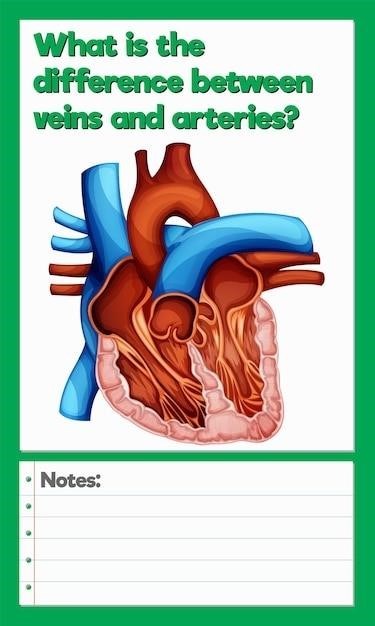
The cardiovascular system, often referred to as the circulatory system, is a complex network responsible for transporting blood throughout the body. It comprises the heart, a powerful muscular pump, and a vast network of blood vessels, including arteries, veins, and capillaries.
The heart acts as the central pump, propelling blood through the arteries, which carry oxygenated blood from the heart to the body’s tissues. Arteries branch into smaller vessels, eventually reaching the capillaries, microscopic vessels where exchange of gases, nutrients, and waste products occurs between blood and surrounding cells. Deoxygenated blood then flows back to the heart through the veins, returning to the lungs for reoxygenation.
The cardiovascular system plays a vital role in maintaining homeostasis, the body’s internal balance. It delivers oxygen and nutrients to cells, removes carbon dioxide and metabolic waste, transports hormones, regulates body temperature, and helps fight infections.
The health of the cardiovascular system is essential for overall well-being. A healthy circulatory system ensures efficient delivery of oxygen and nutrients, facilitating optimal function of organs and tissues. Conversely, problems within the cardiovascular system, such as heart disease or stroke, can have severe consequences for health and longevity.
Understanding the intricate workings of the cardiovascular system is fundamental to recognizing and addressing potential issues, promoting a healthy lifestyle, and preventing or managing cardiovascular diseases.
Common Heart Conditions
While the heart is a remarkably resilient organ, it can be susceptible to various conditions, ranging from minor inconveniences to life-threatening situations. Understanding common heart conditions is crucial for early detection, prevention, and effective management.
One prevalent condition is coronary artery disease (CAD), characterized by the narrowing or blockage of the coronary arteries, which supply blood to the heart muscle. This narrowing often results from plaque buildup, a process known as atherosclerosis. CAD can lead to chest pain (angina), shortness of breath, and even heart attack.

Another common heart condition is heart failure, where the heart muscle weakens, making it less effective at pumping blood. This can lead to fluid buildup in the lungs (pulmonary edema), fatigue, and shortness of breath.
Arrhythmias, or irregular heartbeats, are another prevalent issue. These can range from mild palpitations to potentially life-threatening conditions like ventricular fibrillation. Arrhythmias can be caused by various factors, including heart disease, medications, and lifestyle choices.
Valvular heart disease, characterized by problems with the heart valves, can also significantly impact heart function. The valves may not open or close properly, hindering blood flow and leading to symptoms like fatigue, shortness of breath, and chest pain.
It’s essential to be aware of these common heart conditions and to seek medical attention if you experience any concerning symptoms. Early detection and treatment can significantly improve outcomes and prevent complications.
The heart, a remarkable organ that tirelessly pumps blood throughout the body, is a testament to the intricate workings of the human body. Understanding its anatomy and physiology provides crucial insights into its function and the various conditions that can affect it. From the intricate network of chambers and valves to the coordinated electrical impulses that regulate its rhythm, each aspect plays a vital role in maintaining life.
While the heart is robust, it’s susceptible to various conditions, including coronary artery disease, heart failure, arrhythmias, and valvular heart disease. Awareness of these conditions is crucial for early detection, prevention, and effective management.
By studying the anatomy and physiology of the heart, we gain a deeper appreciation for this vital organ and its importance in maintaining overall health. This knowledge empowers us to make informed decisions about our health and to seek appropriate medical attention when necessary.
The information presented here serves as a starting point for understanding the complexities of the heart. Further exploration through reputable sources like medical textbooks, peer-reviewed journals, and reliable online resources can provide a more comprehensive understanding of this vital organ.

